Anatomy of the eye

We use our eyes in virtually everything we do, and we depend on our vision to steer us through our daily lives. The eye allows us to see the shapes, colors and dimensions of objects by processing the light they reflect or give off. The anatomy of the eye allows humans to see in dim light or bright light, but not in the absence of light. The eye changes light rays into electrical signals and then sends the signals to the brain. The brain interprets these electrical signals as visual images.


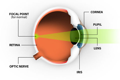
The eye measures approximately one inch in diameter and is set in a protective cone-shaped cavity in the skull called the orbit or socket. The orbit is surrounded by layers of soft, fatty tissue that protect the eye and allow it to turn easily. Six muscles regulate the motion of the eye. Among the more important parts of the anatomy of the human eye are the cornea, conjunctiva, iris, lens, retina, macula and the optic nerve.

The cornea is sometimes referred to as the "window of the eye." It provides most of the focusing power when light enters your eye. The cornea is composed of five layers of tissue. The outer layer is the epithelium. This layer is made up of highly regenerative cells that allows for fast healing of superficial injuries. The two middle layers of the cornea provide strength, shape and structural stability to the eye. The two inside layers work to keep fluid out of the cornea and maintain its optical clarity. LASIK laser vision correction is performed on this part of the eye.

The lens is the clear structure located behind the iris. Its primary function is to provide fine-tuned focusing for near vision. The lens performs this function by altering its shape to become thinner or thicker as necessary. Between the ages of 40 and 50, the lens becomes less flexible and presbyopia sets in. As people reach their 60s or 70s, the lens can become cloudy and hard (cataract formation), preventing light from entering the eye.

The pupil is the hole in the center of your iris (the colored part of your eye). The primary function of the pupil is to control the amount of light entering your eye. When you are in a bright environment, the iris constricts, causing the pupil to become smaller to allow less light to pass through. When it is dark, the pupil expands to allow more light to reach the back of your eye.

The iris provides the color of the eye (i.e. blue/green/brown/hazel). The iris’ main role is to control the size of the pupil through contraction or expansion.

This is the clear, gel-like substance located inside the eye's cavity. The purpose of the vitreous body is to provide a spherical shape to the eye. The vitreous may develop small clumps known as floaters. These are more common in nearsighted people than in the rest of the population.

The retina consists of fine nerve tissue that lines the inside wall of the eye and acts like the film in a camera. Its primary function is to transmit images to the brain. When your vision is perfect, the light rays coming into your eye focus precisely on this part of your eye.

The optic nerve carries electrical signals from the retina to the brain.

The sclera is the white part of the eye’s anatomy. The sclera's purpose is to provide structure, strength and protection to the eye

Common refractive errors

It is important to understand your specific vision problem and how laser vision correction can be used to treat it. Your eye shape and cornea are keys to better vision. When light enters the eye it is bent, or refracted, by a clear tissue on the front of the eye called the cornea. The cornea, in effect, acts like a lens to focus incoming light onto the retina on the back of the eye.


Below are the most common eye problems that can be corrected with LASIK laser correction surgery:

If you have myopia, or nearsightedness, it is because your eye shape is too long and/or you have an excessively steep cornea. As a result of this, light entering your eyes does not focus on the retina as it should, but instead focuses on images at a point in front of the retina. The result of nearsightedness is that your distant objects appear blurry, while near objects appear clear.

If you have hyperopia, or farsightedness, it is because either your eye shape is too short or you have an excessively flat cornea. In farsightedness, light entering your eye focuses on images at a point behind the retina. The result of farsightedness is that near objects you are seeing appear blurry, while distant objects appear clear. In some cases, hyperopia may cause blurriness at both distance and nea.

If you have astigmatism it is because you have a cornea that is not spherical or basketball-shaped as is a normal eye, but your cornea is typically shaped more like a football. The result of astigmatism is that the objects you are viewing are not focused into a single image and vision is distorted or blurry. Astigmatism can be present alone or in addition to nearsightedness or farsightedness.

Remodeling the Cornea:

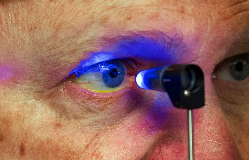
If you are wearing glasses or contacts, these bend the light as needed to adjust for your eye prescription. In a laser vision correction procedure, Dr Sagar Bhargava will use an excimer laser to remodel the cornea to properly refract light for corrected vision. The laser pulses cool, invisible ultraviolet light to painlessly reshape the cornea in a precisely controlled manner without damaging adjacent eye tissue. The cornea then properly refracts light so you can see naturally. These lasers are the most advanced FDA approved equipment to assure you the most precise eye correction available.

Knowing your glass prescription

Knowing your vision prescription is an important part of identifying if you are a good LASIK candidate. You are not required to bring in a previously administered vision prescription; this will be retested during your LASIK evaluation.


After having your eye exam, the doctor will identify the amount of correction needed in each eye. This information will then be used to correct your vision during the LASIK surgery. LASIK laser eye surgery can correct vision impairments such as myopia, hyperopia, and astigmatism. The degree of these conditions is measured in units called diopters. The extent of your nearsightedness, farsightedness, or astigmatism will define your vision prescription in diopter units. This represents the amount of vision correction you need to normalize your eyesight.

  • -7.00 identifies the degree of your nearsightedness or farsightedness. The plus or minus sign identifies whether you are nearsighted (-) or farsighted (+).
  • -2.50 identifies the degree of astigmatism. Sign can be either (-) or (+).
  • 90 identifies where the astigmatism is positioned. This astigmatism is located at ninety degrees, or in the vertical axis.
  • Myopia – Commonly known as nearsightedness . Close objects are able to be seen clearly and objects farther away are blurry. Spectacles wil have – ve power
  • Hyperopia – Commonly known as farsightedness. Objects farther away are clear and objects closer are blurry. Spectacles will have +ve power
  • Astigmatism – Blurred vision most commonly caused by an irregular shaped cornea. Spectacles willhave cylindrical power

Candidate for laser corrective procedure

Before undergoing to your Laser refractive procedure eye surgery evaluation there are some general criteria that can guide you in deciding if this is the right procedure for you. To have laser eye surgery you should be in good eye health, good overall health, and have realistic expectations of the Laser refractive procedure

  • Your eye prescription should fall within certain prescription limits.
  • You should have have eye diseases like keratoconus, glaucoma, cataracts, corneal disease , dry eye and certain retinal and optic nerve diseases.
  • You should have no residual or active eye conditions including optic neuritis, ocular herpes, some cases of amblyopia (lazy eye) AND strabismus (muscle imbalance).
  • You should have no current or recurring infections and ocular allergies.
  • You should have a normal corneal curvature, thickness and pupil size.
  • You must be at least 18 years old.
  • To be a good candidate for Laser refractive procedure, you should not have any autoimmune diseases, such as Lupus, rheumatoid arthritis, or multiple sclerosis.
  • If you have diabetes it must be well managed and under good control and not have diabetic retinopathy.
  • You cannot be pregnant or nursing, or plan to become pregnant in the next 6 months following surgery. If nursing and considering Laser refractive procedure laser eye surgery, you should wait at least 3 months after you’ve stopped. Hormonal fluctuations can affect vision stability.
  • Laser refractive procedure is a highly successful procedure, intended to reduce or eliminate dependence on glasses or contact lenses. The people who are the happiest after Laser refractive procedure are usually those who start out with reliastic expectations about what the procedure can and cannot accomplish. For example, if your goal is to see much better nearly all the time without dependence on glasses or contacts, you are very likely to be very happy with your Laser refractive procedure experience. If your goal is to see even better after Laser refractive procedure than you presently see with your best glasses or contact lenses, you may be expecting more than can routinely be accomplished with Laser refractive procedure.
  • The vast majority of people undergoing Laser refractive procedure will see well enough after the procedure to carry out majority of their activities . Many will see 6/6 , or close to 6/6. However, Laser refractive procedure is not guaranteed to deliver 6/6 vision. Individual results can depend on a number of factors, one of the most important being how your particular eye heals after the procedure.
  • Occasionally, people will find that a weak pair of glasses is still helpful after Laser refractive procedure for certain situations, such as night time driving. It is also possible to experience some starbursts or halos around lights at night after Laser refractive procedure surgery. In most cases these are temporary and not bothersome.
  • It is also important to realize that Laser refractive procedure does not stop age-related changes in the eye. If one continues to become more nearsighted or farsighted after Laser refractive procedure, some increased dependence on glasses may again develop over time. That's why it is best to have Laser refractive procedure when your glasses or contact lens prescription has been fairly stable over time.
  • Laser refractive procedure neither reduces the risk of developing eye conditions later in life, such as cataracts, glaucoma, or macular degeneration nor interferes with the usual treatments for these disorders and Laser refractive procedure does not decrease the risk of certain retinal disorders that are slightly more common in highly nearsighted individuals. So regular complete eye examinations are still important after LASIK.
Pre Refractive surgery Examination

Pre refractive procedure screening

Prior to refractive correction procedure, a comprehensive eye examination to ensure that you are a good candidate for undergoing LASIK/PRK treatment. The tests include:

  • History Taking
  • Visual Acuity Testing
  • Refraction
  • Keratometry
  • Slit Lamp Examination
  • Pupillary Measurements
  • Tear Film Analysis
  • Glaucoma Testing
  • Detailed Retinal Evaluation with Dilatation
  • Oculyzer

Types of laser refractive procedures

1. LASIK (Laser-Assisted in Situ Keratomileusis)

The most commonly performed procedure.
A thin corneal flap is created using a femtosecond laser, lifted, and then the excimer laser reshapes the underlying cornea.
The flap is repositioned after the laser treatment.
Advantages: Quick recovery, minimal discomfort, excellent visual results.

2. Femto-LASIK (All-Laser LASIK)

A bladeless version of LASIK.
Both the flap creation and corneal reshaping are done by lasers, improving precision and safety.
Offers better predictability and fewer flap-related complications.

3. PRK (Photorefractive Keratectomy)

The surface layer (epithelium) of the cornea is gently removed, and an excimer laser reshapes the cornea.
The epithelium grows back naturally over a few days.
Advantages: No flap creation, suitable for thinner corneas.
Disadvantages: Slightly longer healing time and temporary discomfort.

4. SMILE (Small Incision Lenticule Extraction)

A femtosecond laser creates a small lens-shaped piece (lenticule) inside the cornea, which is removed through a tiny incision.
No flap is made.
Advantages: Minimally invasive, faster recovery, less dryness.

  1. Traditional PRK Uses Amoils Brush or a 'Hockey Stick' to remove the corneal epithelium before excimer laser ablation
  2. Alcohol assisted PRK -Uses alcohol to loosen the corneal epithelium before its removal. This allows gentler surgery as it avoids the need for scrubbing or scraping to remove the corneal epithelium. This originated from LASEK, and essentially is LASEK but with removal of the corneal epithelial layer at the end of surgery.
  3. Transepithelial PRK Uses an excimer laser to remove the corneal epithelium. A one step, no touch technique is used where the excimer laser performs both epithelial removal and corneal reshaping sequentially.

The LASIK process also uses the excimer laser to reshape the cornea, but is done under a thin, protective, corneal flap.

Here is a general outline of the procedure:

  1. Anesthetic eye drops are applied to the eye.

  2. The LASIK surgeon creates a protective flap to access the inner corneal tissue. During this part of the procedure, your vision dims and becomes blurry for about a minute. After the flap is created you are able to see the flashing fixation light of the laser and the bright lights used for the procedure.

  3. Next the inner layers of your cornea receive computer-controlled pulses of cool laser light. Although the laser light is invisible, the laser makes a clicking sound as it gently reshapes the inner corneal layer to improve and in many cases, eliminate your prescription. During this part of the procedure, an eye-tracking device tracks your eye movements to ensure precise correction.

  4. Following the re-shaping of the tissue, the LASIK surgeon carefully reposition and aligns the flap to its original position. Protective shields are placed over your eye to prevent accidental rubbing as the flap heals naturally and securely over the next several hours.

Difference between of PRK and LASIK

  PRK LASIK
Pain and discomfort Moderate Mild
Need for bandage contact lens Yes for 3-5 days Not required
Functional visual recovery 1-2 weeks Clear vision from next day
Stable refraction 1 month 2 weeks
Best for Patients with thin cornea and
low errors of refraction
Wider acceptable criteria for correction
Intraoperative complications Nil Flap related complication like free cap , incomplete cap etc
Post operative No major complications except
corneal haze rarly
Flap displacement spontaneous
or trauma related is always a
risk
Good for people who Are into contact sports, combat
situations eg army
Need a very fast visual recovery

Difference Between PRK, LASIK, FemtoLASIK, And SMILE

PRK LASIK FEMTOLASIK SMILE
US FDA approval 1995 1999 2001 2016
Range of correction Up to -6.00 / +4.00 sphere or cylinder Up to -10.00 / +4.00 sphere and -6.00 cylinder Up to -10.00 / +4.00 sphere and -6.00 cylinder -10.00 sphere / -5.00 cylinder
Blade NO YES NO NO
FLAP NO YES YES NO
Corneal thickness >470 microns >500 microns >500 microns >500 microns
Size of incision No cut 20 mm 20 mm 2–4 mm
Intraop complications Nil Flap related Flap & suction loss related Suction loss & lenticule dissection related
Abandonment of procedure No Yes Yes Less chances
Patient cooperation during the procedure Minimal + ++ ++
Post-op discomfort ++ (2–3 days) - - -
Vision improvement 2–4 weeks Next day Next day 1–7 days
Dryness + ++ ++ +
Postop water contact 1 week 2 weeks 2 weeks 1 week
Postop injury No issue Flap displacement Flap displacement No issue
Outdoor sports 4–5 days 2 weeks 2 weeks 3–4 days
Strength of cornea after the procedure ++ - - +
Risk of ectasia 20 per 100,000 eyes (0.020%) 90 per 100,000 eyes (0.090%) — 4.5× higher risk than PRK 90 per 100,000 eyes (0.090%) — 4.5× higher risk than PRK Exact incidence N/A (relatively new procedure)

Magnitude of refractive power that can be corrected

Error Myopia Hyperopia Astigmatism
Range of correction -0.50 D to -12 D Upto +6.00 D Upto 6 D


Procedure PRK LASIK
Central corneal thickness 470 500
Range of correction Upto -6.00 D Upto -12 D

Laser corrective procedure side effects

Laser surgery is very safe and effective. But in some patients there could be side effects.

Sometimes the anterior corneal flap that is made in LASIK may not be complete (called incomplete flap) if the keratome stops mid-way because of suction loss. In this situation the flap is repositioned and ablation is deferred. The surgery is re-attempted after three months. In rare instances, the flap may tear or become detached (called free flap). in this situation your surgeon will decide on table whether to go ahead or defer the surgery. The detached flap can repositioned with a very fine suture.

Dryness in the eyes will persist for 6 months and patients will have to use artificial tear substitute during this period. The symptoms are mainly foreign body sensation , burning sensation and fluctuating vision.

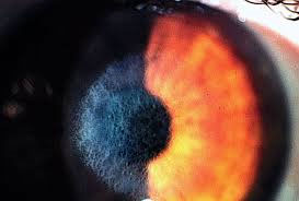
an inflammatory process that involves an accumulation of white blood cells at the interface between the LASIK corneal flap and the underlying stroma. It is known colloquially as "sands of Sahara syndrome" because on slit lamp exam, the inflammatory infiltrate appears similar to waves of sand. It is most commonly treated with steroid eye drops. Sometimes it is necessary for the eye surgeon to lift the flap and manually remove the accumulated cells.

Under correction may sometimes be planned intentionally or may occur as an unintentional effect. As a result, the eye remains short sighted even after the surgery. If the degree of residual myopia is significant, the eye may be retreated at a later date. Over correction can occur very rarely.

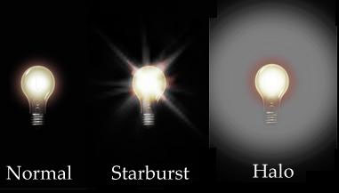
You may feel some sensitivity to light at night or in bright sunlight. Sometimes in dim light, you may see a faded ghost image around the sharp bright image. This will pass after the first few days or weeks. In rare circumstances this may remain on long term.

Some people find that their night time vision has become a bit dull. This happens because of a decrease in their ability to discriminate between different contrast levels.

In some rare cases, there can be development of corneal haze after treatment. Now a days Mitomycin C application is done during the procedure which has helped in reducing the haze incidence.

It is a very complication of laser correction procedure. It can occur if the corneal thickness is less to begin with, or if the cornea is thinned more than it can withstand with the lasers. Therefore, persons having inadequate corneal thickness are not suitable candidates for LASIK.

Serious complications like corneal infections though possible, are extremely rare.

FAQ's

Wavefront-optimized LASIK is the type of laser treatment available on the Wavelight laser. This treatment is also based On the patient’s eye power and also takes into account corneal curvature and thickness, and applies laser energy in a unique fashion in the periphery of the cornea. This laser has been found to reduce the aforementioned complications such as glare, halos, and other nighttime visual aberrations that can occasionally occur with conventional treatments.

You should be able to see reasonably well within a few days after your procedure. If you have had a SURFACE PRK procedure, your vision may fluctuate for the next 1 to 3 months and reading fine print may be difficult. If you have had a LASIK procedure, your vision is likely to stabilize around a month after the procedure.

Early side effects of any corneal procedure include light sensitivity, glare and foreign body sensation in the eye. Both SURFACE PRK and LASIK can sometimes result in under correction or incomplete removal of the refractive error. Occasionally, an overcorrect ion eg., the correction of more myopia than intended, can also occur. A change in the clarity of the corneal tissues – termed corneal haze – can also occur in some patients.

A large percentage of nearsighted, farsighted, and astigmatic patients are potential candidates for LASIK. Patients who are 18 years of age or older, have healthy eyes that are free from retinal problems, corneal scars, and any eye disease are suitable. Along with being medically suitable, the best candidates are people with a lifestyle or occupation in which they are dissatisfied with their contact lenses or glasses. By having an evaluation, examination, and consultation with you, the doctor will be able to determine if you are a good candidate.

In our practice, LASIK is performed as an office procedure in the comfort and convenience of an excimer laser suite. The entire treatment takes about 5 to 7 minutes per eye or approximately 15 minutes for both eyes.

Yes. Following an initial healing period of two to three months, the effect of the treatment is lifelong.

Generally, patients under 40 years of age read well without glasses following the treatment. Patients over 40 may need magnification for reading fine print. Presbyopia is the term that refers to the natural weakening of the focusing muscles that occurs in our early to mid 40s, causing us to need the magnification that reading glasses provide. The LASIK treatment does not correct or prevent presbyopia.

Your physician will advise you as to the specifics, however, typically you can bathe the same day of the treatment but you should not shower for 2 weeks after the treatment. As always, avoid getting soap or water directly into your eyes. Avoid rubbing your eyes during the first month after surgery.

You should not drive on the day of the treatment. After that, you may drive when you feel confident that your visual acuity and eye comfort allow you to drive safely.

You may resume wearing eye makeup 4 weeks after your treatment. It is strongly recommend that you purchase new eye makeup, specifically mascara, to avoid potential infection following your treatment.

Non-contact sports can be resumed as soon as you feel capable. Eye protection is always recommended for racquet sports or rough sports where there is a risk of being hit in the eye.

You should stay out of the swimming pools for two weeks after your treatment, and rivers, lakes and oceans for 3-4 weeks following your treatment.

Numbing eye drops are used. Sometimes an oral medication is used to help with relaxation. No needle or intravenous drugs are used.

In LASIK– Both eyes are performed same time, but SURFACE PRK and Epi LASIK is performed one eye at a time. The timing of treatment of the second eye is best decided in consultation with your ophthalmologist.

The treatment itself is painless because we put a few numbing drops in your eyes to make you even more comfortable. You will feel pressure on and around the eye during treatment. With LASIK, some patients may experience a feeling of "something in the eye" for a day or so after treatment.

You will be "awake" but very relaxed. You will not be able to see any of the details of the treatment. You will see a number of lights, but the images will be blurred.

Some patients experience a feeling of burning or scratchiness. This feeling generally lasts from one to three hours following the treatment. Most people feel fine if they take a nap soon after the treatment.

No, but clear, protective eye shields will be placed over your treated eye(s) following the treatment, and will be removed in our office the next morning. You will continue to wear these eye shields at bedtime for 1 week for protection.

Both SURFACE PRK and LASIK are designed to allow you to discontinue the use of corrective lenses. In some patients with high refractive errors and in those over 40 years of age, corrective lenses may still be required. In some patients however, it may be possible to perform a second refractive procedure to correct a residual refractive error.

LASIK (Corneal Refractive Surgery) is considered as elective/ cosmetic surgery and it is not covered by many health insurance plans. You may also contact your insurance agency for the same.

If you have had a SURFACE PRK, you will be able to return to work within three to four days of surgery. If you have had LASIK procedures, you can return to work on the following day. Eye medications should be continued during working hours as instructed by the surgeon. While most activities can be resumed after this time, the face should not be placed under water for at least one week following LASIK. Care should also be taken to avoid any trauma to the eye including rubbing of the eyes during this period.